
Anatomy of the Eye
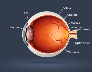
- Retina
The thin lining at the back of the eye that is responsible for vision. The rods and cones at the back of the eye convert light to the optic nerve then to the brain for analysis. - Macula
The central portion of the retina responsible for the sharp, detailed vision. - Vitreous
The clear jelly-like substance that fills the rear two-thirds of the eye. The vitreous helps filter light to the retina as well as maintain the structure of the eye as a whole. - Sclera
The outer coat of the eyeball that forms the visible white of the eye and surrounds the optic nerve at the back of the eyeball. - Choroid
Layers of blood vessels located between the sclera and the retina; they provide nourishment to the back area of the eye. - Fovea
A depression in the retina that contains only cones (not rods), and that provides acute eyesight. - Optic Nerve
The nerve that carries electrical impulses from photoreceptor cells (rods and cones) in the retina to the visual cortex in the brain. - Lens
The nearly spherical body in the eye, located behind the cornea, that focuses light rays onto the retina. - Cornea
The clear part of the eye covering the iris and pupil; it lets light into the eye, permitting sight.